Coma: Functional MRI to predict first signs of awareness
Terry Shivo was 27 when she collapsed. Paramedics found neither respiration nor a pulse. Almost three months later her eyes had opened, but she was not conscious. She has a sleep/wake circle and sometimes appeared to smile - she was in a vegetative state.

Up to her death in the USA in 2005 her case caused a bitter court battle for guardianship between family members and a worldwide controversial debate began about the quality of life following severe brain damage.
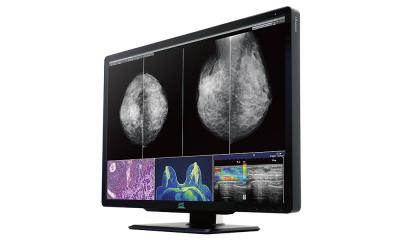
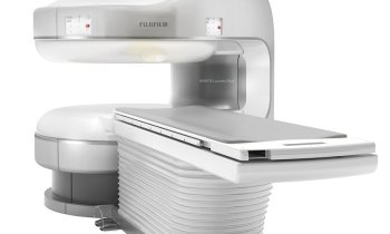
Reliable methods of diagnosis could give more certainty about signs of awareness and chances of recovery. Presently, the Functional Magnetic Resonance Imaging (fMRI) is the most promising tool not only to provide better diagnosis and predict outcome of brain injuries but also to communicate with the patient.
In Europe only two research centres perform this imaging technique on coma patients: the Cambridge Neuroscience Centre in the UK and the Coma Science Group from the Cyclotron Research Centre at the University of Liège, Belgium. The latter is led by Professor Steven Laureys, Head of Clinics in the Department of Neurology.
Asked why coma research is so important, the professor said that, due to modern emergency medicine and intensive care, an increasing number of patients survive injuries, toxications or hypoxia.
‘Unfortunately they are often considered as a homogenous group of hopeless cases, but this is not true. Studies have proved that around three or four in 10 patients diagnosed as unconscious in fact show signs of awareness. There are at least four different states of severe brain damage we observe: coma, vegetative state, minimally conscious state (clearly discernible evidence of consciousness of self or environment) and locked-in-syndrome (fully conscious but paralysed). Especially the demilitation of vegetative and minimally conscious states is very difficult to differentiate. fMRI is helping us to understand the brain function and cognitive processes.
‘Functional magnetic resonance imaging (fMRI) shows the function of the brain in contrast to MRI which displays structural images. The haemoglobin in the red blood cells transport oxygen to the brain and can be used to make scans with the help of a magnet field. So actually, we measure the blood oxygen level, also called haemodynamics, indirectly to obtain information about the activity of neurons in special parts of the brain. But fMRI can do more: The technique enables us to communicate with the patient. We can ask him questions by telling him to think of playing tennis if he wants to say “yes”, or imaging to walk through his home if he wants to say “no”. We then directly see the reaction of brain activity in specific areas on the monitor. It permit us to give back autonomy to the person to decide for himself if, for example, he wants life-prolonging measures.’
‘In functional neurological imaging fMRI replaces PET scanning, which requires radiotracers injection. With fMRI you can possibly acquire more scans and a better resolution in time and space. According to time, tools to measure electrical activity from the scalp are even more precise – these would be EEG and Magneto-encephalography (MEG). That’s why the gold standard is to record simultaneously both: EEG during fMRI. The present challenge is that MRI needs a very high magnetic field – at the moment 3-Tesla, but we can move up to 7T, or even higher.
Unfortunately the signals of both tools distort each other. So the EEG electrodes must be shielded to avoid image interference and burning at the patient.
Presently we have no direct treatments to help the brain recover from traumatic or anoxic cases, because first we would have to know what gives rise to recovery of consciousness. We know that the vegetative state can be permanent or transitory and that the longer someone stays unconscious the smaller their chance of recovery. Retrospective studies indicate that patients taking specific drugs apparently recover more often. In the future, fMRI could help to measure the effects of drugs testing directly to the central nervous system.
01.09.2008