XPCi
New technique enables transfer of X-ray phase contrast imaging to clinical practice
By Professor Robert D Speller, Head of the Radiation Physics Group, University College London, and Dr Alessandro Olivo, of the Medical Physics & Bioengineering Dept. University College London
X-ray phase contrast imaging (XPCi) is a novel imaging technique with the potential to revolutionise the field of diagnostic radiology. It can do this because it is based on a different physical effect, namely refraction/interference instead of absorption. Studies carried out with synchrotron radiation have demonstrated that the exploitation of such effects leads to a substantial increase of the contrast of all details in an image, as well as to the detection of details classically considered invisible. These studies have shown impressive advantages especially in those fields where small absorption differences are the main factor limiting image quality, such as mammography – where most tumours are characterised by X-ray absorption characteristics very similar to the ones of the surrounding healthy tissue. After preliminary investigations based on ex-vivo studies, which demonstrated substantial improvements both in terms of sensitivity and specificity, the first station for in vivo XPCi mammography with synchrotron radiation is currently in operation in Trieste, Italy. Alongside mammography, substantial benefits in many other radiology fields were demonstrated by means of ex-vivo or animal studies. These include:
- lung imaging, in which the technique showed the potential to spot small lesions with conventional planar imaging without having to rely on expensive (both in terms of cost and patient dose) CT scans
- vascular imaging/coronary angiography, in which the potential to image blood vessels without contrast agents was demonstrated
- bone imaging, where minimal details on the bone trabecular structure are easily and effectively depicted due to the substantially increased sensitivity
- and many others, to include improved resolution and lesion detectability in liver imaging, kidney imaging, etc. Moreover, refraction/interference effects are less subject to decreasing with increasing X-ray energy that absorption effects: as a consequence, images could be acquired at higher X-ray energies, which could translate into dose reductions also of one order of magnitude.
Despite being probably the ideal X-ray imaging technique, the problem so far with XPCi was that its use seemed to be restricted to synchrotron radiation environments. All early implementations of the technique seemed in fact to require levels of spatial coherence (i.e. small focal spot plus large source-to-sample distance) and monochromaticity not available with state-of-the-art clinical sources. Although pilot experiments with synchrotron radiation like the one on mammography currently underway in Trieste have a high scientific significance, a real world-scale impact would be achieved only by developing a relatively small-sized, cost effective prototype. This clearly requires taking XPCi out of synchrotron environments and into laboratory practice.
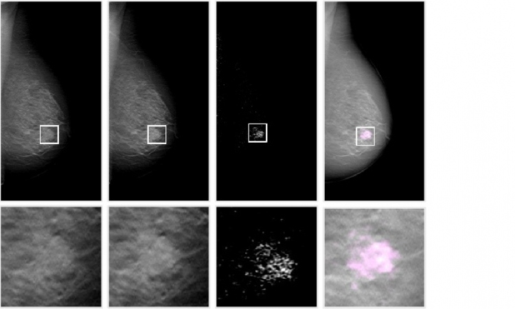
Researchers at University College London, after having demonstrated that polychromatic radiation can provide the same level of phase contrast image quality of its monochromatic counterpart, have developed a new technique based on the use of coded apertures, which makes all advantages of XPCi achievable with conventional sources.
Unlike other techniques based on perfect crystals, grating interferometers, etc, the coded apertures approach allows for the first time the use of divergent, polychromatic X-ray beams like those produced by conventional clinical sources. Moreover, the combined effect of two sets of apertures strongly relaxes the requirements on the source coherence, and it was demonstrated that levels of phase contrast signals comparable to the ones obtained with synchrotron radiation can now be achieved with source sizes as big as 100 hm – i.e. fully compatible with current mammography sources. This technique has therefore the potential to allow for the first time an effective transfer of XPCi into clinical practice, and a consequently widespread diffusion of the technique.
The new technique was fully modelled through a computer simulation, a small imaging prototype was realised, and the pilot experiments carried out provided results perfectly matching the simulated ones. Such experimentation confirmed beyond doubt that the advantages of XPCi demonstrated by synchrotron radiation studies can now be achieved by means of conventional X-ray sources.
A system based on the new technique would therefore be based around conventional, state-of-the-art sources and detectors. As a consequence, its cost would not differ substantially from radiography units currently in use.
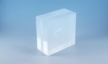
The main difference would lay in the introduction of the coded-aperture arrays, which consist of extremely thin (20-30mm) gold layers deposited on graphite substrates. The cost of such a device is currently in the range of a few thousand Euros, but could be reduced by at least one order of magnitude if the devices were mass-produced. Alignment stages to achieve the correct positioning of such devices would be the only further addition to the system, meaning that its practical realisation would thus be very cost-effective.
Contact: aolivo@medphys.ucl.ac.uk
01.03.2008