Image source: Washington University in St. Louis/Zhu lab
News • Tissue oxygenation
Photoacoustic imaging for better diagnoses of ovarian cancer
Ovarian cancer is the deadliest cancer of the female reproductive system, and there is no screening test that can help with early detection. Ultrasound imaging, the standard of care used to determine whether lesions are cancerous or benign, is not always accurate, leading some patients to have the ovaries removed unnecessarily.
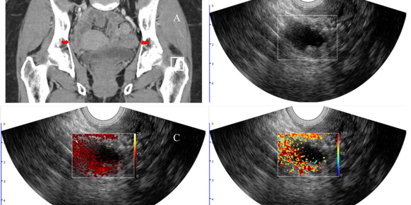
Researchers and clinicians at Washington University in St. Louis have found a way to improve the standard of care diagnostic accuracy of potentially cancerous lesions in the ovaries and adnexal regions, or the fallopian tubes, by incorporating functional biomarkers with photoacoustic imaging, a technique that illuminates tissue with near-infrared light at specific wavelengths that are absorbed differently by oxygenated and deoxygenated hemoglobin.
Results of this new additional assessment technology may allow some patients to avoid surgical removal of abnormally appearing lesions, reducing health care costs and potential complications from the procedure. The findings of the research were published in Ultrasound in Obstetrics and Gynecology.

Image source: Washington University in St. Louis
Quing Zhu, the Edwin H. Murty Professor of Engineering at the McKelvey School of Engineering, worked with a team of Washington University School of Medicine physicians, led by Matthew Powell, MD, director of the Division of Gynecologic Oncology and a professor of obstetrics and gynecology; and Cary Siegel, MD, a professor of radiology and chief of the gastrointestinal/genitourinary fluoroscopy section in the Mallinckrodt Institute of Radiology, to add photoacoustic imaging to the standard of care for women scheduled to have their ovaries and/or adnexal lesions surgically removed due to potential malignancy.
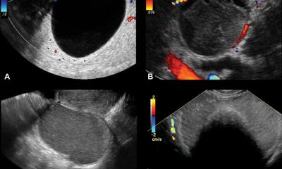
Photoacoustic imaging has been used to assist in diagnosis of several other cancers, including breast, skin, colorectal and other cancers of the female reproductive system; however, it has not been used to classify adnexal malignancy risk, the researchers said. “Photoacoustic imaging combined with ultrasound provides complementary diagnostic imaging data involving structure and function,” Zhu said. “The ultrasound localizes the lesion, and the photoacoustic images inform tumor hemoglobin content and percent of blood oxygen saturation.”
Hemoglobin concentration and blood oxygen saturation provide functional information on the vasculature within the tissue as well as oxygen consumption. Malignant lesions generally have higher hemoglobin concentration due to tumor angiogenesis and lower blood oxygen saturation due to high tumor metabolism, a finding that helps differentiate benign from malignant lesions.
Women frequently develop ovarian cysts, which cause a great deal of worry. This technology should quickly and easily be able to help assure both the doctor and the patient that most of these can be watched, and unnecessary surgery can be avoided
Matthew Powell
In a clinical study conducted by the team, 68 patients scheduled to have their ovaries surgically removed underwent clinical ultrasound as well as the combined photoacoustic and ultrasound technology (PAT/US) that Zhu’s team developed. Among the patients, 14 had malignant lesions in their ovaries or adnexal regions, two patients had malignant fallopian tubes and 52 patients had benign lesions. Each lesion was graded by two radiologists using the Ovarian-Adnexal Reporting and Data System (O-RADS), which classifies risk for malignancy in six categories, from O-RADS 0 being an incomplete evaluation, to O-RADS 5, meaning a more than 50% risk of malignancy.
“Women frequently develop ovarian cysts, which cause a great deal of worry,” Powell said. “This technology should quickly and easily be able to help assure both the doctor and the patient that most of these can be watched, and unnecessary surgery can be avoided. These surgeries can lead to loss of fertility and early menopause. This technology and other imaging advancements could be immensely helpful to improve the lives of our patients.”
When comparing the malignant and benign lesions, the relative total hemoglobin in the patients with malignant lesions in the ovaries and in the adnexal region was 1.8 times higher than in those with benign lesions. The blood oxygen saturation was 5% lower in those with malignant lesions, though it was not a statistically significant difference. Presence of the cancer antigen CA 125 was significantly different in patients with malignant lesions compared with patients with three common types of benign lesions but not significantly different from the group with endometriosis, abscess, adhesions, infarction or torsion.
Ultimately, the team found that the most important predictors of malignancy were relative total hemoglobin, the O-RADS score, presence of the cancer antigen CA 125 and percentage of blood oxygen saturation (sO2). Their model that incorporated all four features had an area under the receiving characteristic curve (AUC) of 0.97, a metric used in medical imaging diagnosis to measure a model’s performance. A score of 1 indicates ideal model performance.
“The assessment of the adnexal mass may be challenging for an experienced radiologist, as many different benign and malignant causes are often in the possibilities for diagnosis,” Siegel said. “Novel imaging techniques like photoacoustic ultrasound that increase the radiologist’s accuracy and confidence are critical to improving early diagnosis and treatment of ovarian cancer. With early diagnosis, our ultimate goal is to improve ovarian cancer survival. Working with Dr. Powell, Dr. Zhu and her engineering team has been a highlight of my medical career.”
Source: Washington University in St. Louis
09.12.2023