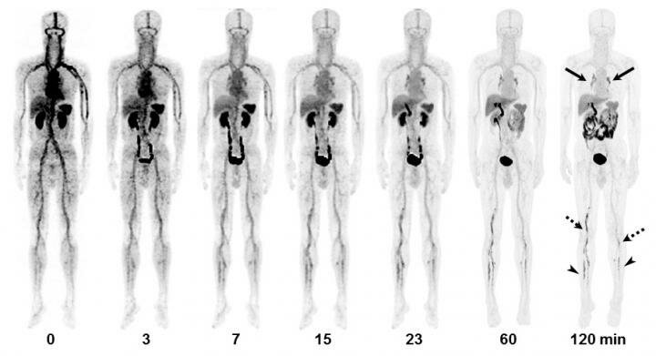
Credit: Kim C, Lee JS, Han Y, et al.
News • Imaging agent
PET/CT tracer offers better diagnosis of acute venous thromboembolism
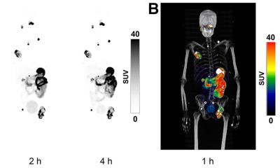
A first-in-human study reports that the novel positron emission tomography/computed tomography (PET/CT) tracer 18F-GP1 showed excellent image quality and a high detection rate for the diagnosis of acute venous thromboembolism (VTE). Well-tolerated in patients, 18F-GP1 PET/CT also identified blood clots in distal veins of the leg below the knee, where conventional imaging has limitations.
Acute VTE is a disease that includes deep-vein thrombosis of the leg or pelvis and its complication, pulmonary embolism—which can be fatal. The highly variable and nonspecific symptoms and signs of VTE often result in delayed or inaccurate diagnosis. For acute VTE, timely and accurate diagnosis is critical to expedite the initiation of effective therapeutic strategy. "Conventional imaging with ultrasonography, CT venography or CT pulmonary angiography is typically unable to distinguish old thromboemboli from new and potentially unstable thromboemboli," stated Dae Hyuk Moon, MD, professor at Asan Medical Center, University of Ulsan College of Medicine in the Republic of Korea. "The 18F-GP1 tracer used in this study offers the unique ability to detect, characterize and track newly formed thrombi that have a high risk for embolization and further complication."
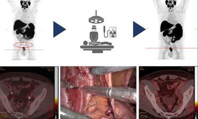
Researchers conducted a prospective study to obtain clinical proof-of-concept for thrombus PET imaging with 18F-GP1. The safety and diagnostic performance of 18F-GP1 PET/CT were assessed in 20 patients with acute deep-vein thrombosis or pulmonary embolism (10 deep-vein thrombosis and 10 pulmonary embolism). Each patient had signs or symptoms of VTE and had one or more VTE foci confirmed by standard imaging.
Upon image review, researchers found that 18F-GP1 uptake in thromboemboli was easily distinguishable from the blood pool. Moreover, a positive correlation was observed between 18F-GP1 uptake and P-selectin expression on circulating platelets, which shows the presence of activated platelets and acute VTE. 18F-GP1 PET/CT detected thromboembolic foci in all 20 patients with deep-vein thrombosis or pulmonary embolism. Additionally, 18F-GP1 PET/CT showed an increased uptake in the distal veins of the leg in 12 patients that was not detected with conventional imaging. "Incorrect diagnosis of VTE commits the patient to unnecessary anticoagulation and results in higher risk and costs, whereas incorrectly concluding that VTE is absent places the patient at high risk of potentially fatal pulmonary embolism," said Moon. "Although the current studies are preliminary, 18F-GP1 PET/CT may provide not only more accurate anatomic localization, but also information on the risk of the clot growth or embolization. This may lead to changes in clinical intervention to the individual patient."
Source: Society of Nuclear Medicine and Molecular Imaging
18.02.2019