Proven Benefits of ShearWave Elastography
Radiology and European Radiology Journals Report High Reproducibility and Significant Improvement of Breast Ultrasound Specificity. The much anticipated results of the largest clinical breast study ever sponsored by an ultrasound manufacturer have recently been published in the prestigious peer reviewed Radiology and European Radiology medical journals.

The multinational, multicenter “Breast Elastography 1 (BE1)” study, involving renowned clinicians in the breast radiology community, assessed the clinical benefits of ShearWave Elastography in the ultrasonic diagnosis of breast lesions.
The study had two objectives: The first was to demonstrate that images obtained using ShearWave Elastography were reproducible. The second was to compare ultrasound alone versus the combination of ultrasound and ShearWave Elastography for breast lesion characterization. The goal of the latter was to improve lesion classification in categories BI-RADS 3 and BI-RADS 4, where most treatment dilemmas occur, in order to better direct patients towards clinical shortterm
follow-up or biopsy.
The BE1 multicenter clinical trial
A multinational, multicenter breast clinical study was launched in April 2008 with 17 prestigious American and European sites including: the Hammersmith Hospital, Imperial College of Medicine, London (United Kingdom), the Curie Institute of Paris (France), the DKD Wiesbaden and the academic hospitals in Greifswald and Kiel, Schleswig-Holstein (Germany), Yale Medical Center and the Northwestern Memorial Hospital, Chicago (USA). The study was conducted under the leadership of Professor David Cosgrove (Imperial College of Medicine, London) and over 1800 breast lesions were assessed. Clinical Result 1: ShearWave Elastography features were accurate and reproducible1.
To determine reproducibility, each clinical investigator was asked to perform and compare features on 3 separate ShearWave Elastography image acquisitions of the same lesion. The clinical results clearly showed that ShearWave Elastography was reproducible both qualitatively and quantitatively:
• Qualitative: 88% of consecutive repeated ShearWave Elastography exams were rated by physicians to be all 3 similar in appearance.
• Quantitative*: ICC2 rates for ShearWave Elastography measurements were close to perfect, ranging from 0.84 to 0.95
The reproducibility of any imaging modality assures the physician of a reliable and precise evaluation of a lesion, both during the examination and over time. Reproducibility is key for reporting, follow up and therapy monitoring. Clinical Result 2: ShearWave Elastography increased specificity and Positive Predictive Value (PPV) values. 3: Using the ShearWave Elastography Maximum Value (kilopascals), ultrasound specificity increased by 26% with no loss in sensitivity and the PPV for biopsy of the BI-RADS® 4a class increased by 122%. Using the ShearWave Elastography Color-Coding of Maximum Stiffness, the specificity of ultrasound diagnosis increased by 28% with no loss in sensitivity and the PPV for biopsy of the BI-RADS® 4a class increased by 155%.
Claude Cohen-Bacrie, Executive Vice-President of SuperSonic Imagine and Senior Author of the two publications states:
“The outcome of this study goes well beyond our expectations. The study protocol, as it was designed, did not introduce any a priori on the role of SWE as a complement to B-Mode. These published findings are providing the groundwork for a major clinical breakthrough in breast ultrasound. The results will certainly have an important impact on patient management in an area where the need for improvement in screening, diagnosis and therapy has been a driver for the entire medical community.
As a medical imaging company, SuperSonic Imagine has always demonstrated a strong commitment to clinical research. This study, conducted with the best-known breast ultrasound experts, is a perfect illustration of our devotion to finding answers to difficult clinical questions.”
ShearWave Elastography: The Technology
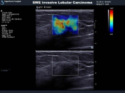
ShearWave Elastography is a technological achievement that gives additional, important quantitative information about tissue elasticity to ultrasound imaging. Unlike conventional elastography methods, which rely on manual compression and measure tissue displacement, ShearWave Elastography requires no manual compression and computes true tissue elasticity by measuring the velocity of shear waves as they propagate in tissue. Shear wave propagation speed in tissue is directly related to tissue stiffness.
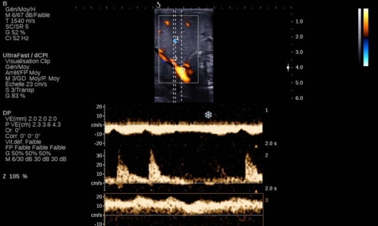
This technology relies upon the generation of a shear wave and its subsequent capture, only made possible with UltraFast Imaging. UltraFast Imaging acquires images at up to 20,000 Hz, 200 times faster than conventional ultrasound. Shear wave propagation speed is calculated and a color-coded, real-time ShearWave Elastography map is produced showing quantitative (kilopascals), local tissue stiffness.
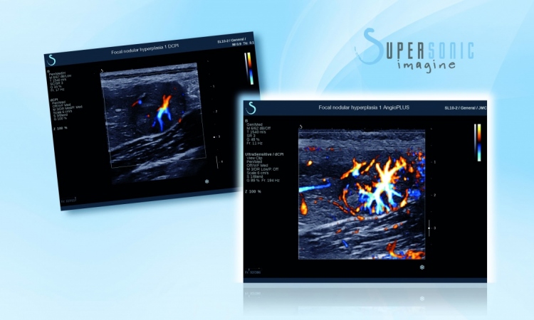
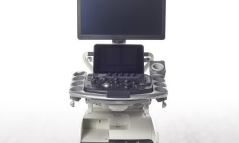
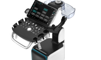
ShearWave Elastography has been used in various organs including the liver, prostate, thyroid and breast with promising and compelling results and it is only available on the Aixplorer MultiWave Ultrasound System.
####
1
ShearWave Elastography for breast masses is highly reproducible. Cosgrove DO et al. European Radiology
2011 Dec 31 http://www.springer.com
2ICC Intraclass Correlations Coefficient
3 Shear-wave Elastography Improves the Specificity of Breast US: The BE1 Multinational Study of 939 Masses
Radiology (2012 Feb;262(2):435-449) by Wendie A. Berg, MD, PhD, et al. © RSNA, 2012 - http://radiology.rsna.org/content/262/2/435.abstract
* The Aixplorer System legally available for sale in the U.S. does not include the quantification tool.
04.03.2012