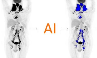
Hybrid imaging: PET-CT and MR-PET
A series of papers presented at the European Congress of Radiology on Friday have highlighted how hybrid imaging is helping radiologists achieve better results in the diagnosis of patients’ conditions. In a session focussing on molecular imaging and entitled “Hybrid imaging: PET-CT and MR-PET”, findings from ten different research papers were detailed by radiologists from Italy, Switzerland, Germany, Japan, India and Saudi Arabia.


Dr Kazuhiro Kitajima from the Department of Radiology at Kobe University and his team compared low-dose non-enhanced CT with full-dose contrast-enhanced CT in integrated FDG-PET/CT studies for the diagnosis of ovarian cancer and they discovered that the PET contrast enhanced CT is an accurate modality for assessment of ovarian cancer recurrence.
A team from Padua, Italy, found that PET-CT was more accurate than CT in the detection of bone marrow metastases while researchers from the University of Tubingen, Germany, showed that contrast-enhanced CT improved malignant lesion classification. Dr Paoletta Mirk, from the Department of Radiology at the Catholic University of the Sacred Heart in Rome, outlined her team’s work on the “additional value of dual-phase 18F-FDG PET-CT in recurrent gynaecological malignancies.” They compared it with standard PET-CT and found it had higher sensitivity and higher accuracy. She said: “Dual phase PET-CT seems to improve diagnostic accuracy for patients with gynaecological malignancies.”
A team from Saudi Arabia found FDG PET-CT was a useful tool in the follow up of paediatric patients with Ewing sarcoma and primitive neuroectodermal cancer with high diagnostic accuracy. Dr Till Heusner from the Department of Diagnostic and Interventional Radiology at the University Hospital Dusseldorf, carried out work with a team to assess the diagnostic accuracy of virtual FDG-PET/CT bronchoscopy for the detection of lymph node metastases in non-small cell lung cancer patients and found it “technically feasible, gave relatively high diagnostic accuracy, and gave access to the bronchi even in the periphery of the lung.”
Dr Katja Pinker-Domenig presented work from the Medical University Vienna on molecular imaging of breast tumours with MR-PET and talked of the benefits to radiologists and patients of fusion of imaging modalities.
She said: “Multi-parametric PET-MRI is feasible, accurate and obviates unnecessary breast biopsies for patients by 50%.”
The session was moderated by Professor Katrine Riklund from Umea University in Sweden and Dr Letterio Politi from the San Raffaele Scientific Institute in Milan. By Mark Nicholls.
03.03.2012