11.7-T scanner a magnet to draw international researchers to France
A massive scanner for in vivo molecular imaging of the human brain will reveal the metabolic processes of neurological disorders, John Brosky reports
Sometimes big science is needed to answer the big questions. It took, for example, the ambitious Hubble telescope to provide new insights into the vastness of the universe, while the Large Hadron Collider (LHC) near Geneva, Switzerland is expected to reveal the deepest secrets of the smallest known particles.
Now a massive Franco-German project under construction outside of Paris is targeting another mysterious zone that has escaped our understanding, the human brain.
Scientists and doctors have penetrated and probed the brain for centuries, yet these explorations have not yielded an explanation as to how it works or why things can go wrong.
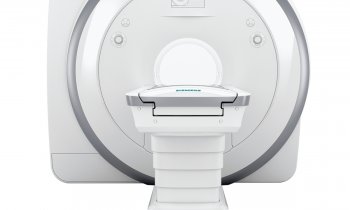
NeuroSpin, the 11,400 meter-square complex in Saclay, France, is already equipped with two magnetic resonance imagine (MRI) machines of 3-tesla and 7-tesla, but in 2012 it will take delivery of a the world’s most powerful MRI magnet dedicated to human medical research -- a wide bore, whole body scanner of 11.7-T.
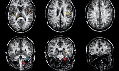
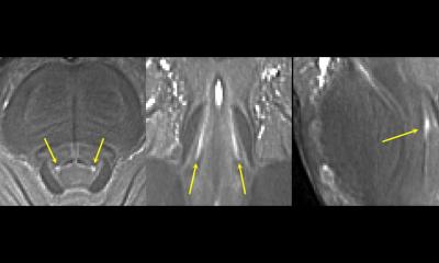
Where the typical MRI scanner of 1.5-T used in hospitals can provide images of anatomy, the Neurospin MRI, at eight times greater sensitivity, will offer both functional views, to study cognitive processes in the normal and diseased brain, and molecular imaging, to watch the workings of the cells.
According to Denis Le Bihan, the project’s initiator and scientific director of NeuroSpin, the goal of the massive centre is to ‘push back the existing limits of brain imaging to obtain accurate images of as little as a tenth of a millimetre, that is to say the size of a group of neurons.’
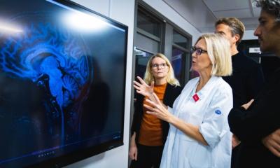
Inspired by the collaborative research model used for the Hubble telescope or the LHC, the Neurospin complex has been conceived as a shared facility designed to welcome international research teams to carry out their studies.
Neurospin will also house a fully equipped clinical wing with hospital beds and examination rooms to accommodate patients and participating study volunteers.
The pan-European partnership coordinating this research believes in vivo molecular imaging of the human brain is the key to unlocking our understanding of the metabolic processes of neurological disorders, such as Alzheimer’s’ or Parkinson’s disease.
Professor Richard Frackowiak, director of Cognitive studies at France’s Ecole Normale Supérieure explained: ‘Early detection of a disease such as Parkinson’s is critical because once a person exhibits the first signs of the disease they have already lost 70% of the neurons specific to this disease. If we can detect this pathology at a stage where only 20% of the neurons have degenerated, the hope for slowing the disease, and beyond that, the survival of the patient becomes possible.’
Each year developed countries are recording a growing percentage of the population suffering neurological or psychiatric diseases and new tools and remedies would have a direct impact on healthcare across a range of activities, including medical imaging, neurosurgery, electrophysiology or psychiatry.
Citing the long range benefit of an improvement of public health through early detection and follow up of neurological pathologies, and noting ‘there are no immediate market incentives for a programme of this nature,’ in December 2008 the European Commission gave its approval for a €54 million investment from France for an upstream development programme called Iseult, which is based at Neurospin.
The Iseult consortium of manufacturing groups and public research laboratories is being led by Guerbet (France) the developer of medical imaging contrast agents, and includes Siemens (Germany), Bruker (Germany), the Atomic Energy Commission (CEA)of France and the University of Freiburg (Germany).
The estimated total cost of the programme over eight years is €137 million, of which the French partners are contributing €109 million.
The Iseult project is focused on three converging research paths to solve the daunting challenge of first building the 11.7-T scanner, and then the not less formidable difficulties of developing contrast agents and software to render images on a computer monitor that scientists and medical researchers can see and interpret.
Guerbet will develop new contrast products for MRI molecular imaging adapted for high field magnetic applications destined for early diagnosis and therapeutic monitoring of Alzheimer's disease, cerebrovascular accidents and brain tumours.
Siemens, the manufacturer of the two MRI scanners already operating at Neurospin, will develop the specific instrumentation and software tools for image analysis required to use the contrast products developed by Guerbet under the Iseult programme.
Siemens is also working with the considerable resources and expertise of the CEA to construct the ultra high-field whole body scanner. Neurospin is located within the sprawling CEA complex at Saclay.
At 11.75-T, the Neurospin MRI is reaching the physical limit at which it is possible to use established technologies for MRI machines, such as cooling of the coils, according to Pierre Védrine, CEA’s director of accelerators, cryogenics and magnetics programmes.
Another sizeable difficulty is the magnet itself, which, for a 90 cm bore to accommodate whole body scans, needs more than 100 tons of equipment and 136 kilometres of cables and wiring, he pointed out: ‘This is a real brain-teaser in terms of electromagnetic constraints.’
12.11.2009