Realtime-Sonoelastography to detect and characterize breast lesions
New possibilities in breast ultrasound comprise techniques for optimizing image quality such as tissue harmonic imaging (THI) and frequency compounding (FC), tools for post-processing US raw data such as strain imaging (elastography), and the use of Realtime-Sonoelastography, PD Dr. Anke Thomas from the Charité in Berlin, Germany, introduced her speech at the Hitachi Symposium Realtime Tissue Elastography Proven diagnostic performance at this years ECR. During her speech, she focused on characteristic features of malignant breast lesions using these techniques with special regard on the sonoelastography.
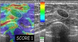
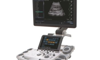
Realtime Tissue Elastography bases on the conventional B-mode image and can easily be integrated into the routine ultrasound examination: strain images are generated in real-time with the Extendes Combined Autocorrelation Method, using a freehand approach to apply gentle compression.
The strain image is calculated and displayed as a colour overlay on the conventional B-mode image, the more stiff tissue structures being displayed as blue whilst the more easily deformed tissues are displayed as red.
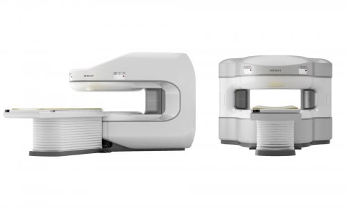
Hitachi Medical Systems already offers the second generation of its Real-time Tissue Elastography (HI-RTE) and it has been proven in a variety of different clinical areas like breast and urology.
In breast applications, HI-RTE has a complementary diagnostic role to the conventional B-mode: Whereas the accuracy of conventional ultrasound is directly related to size, the technology demonstrates a high accuracy in breast lesions smaller than 2 cm. Moreover it increases the specificity of conventional ultrasound especially in masses categorised as BIRADS 3 and 4, adding new benign criteria and therefore reduce unnecessary procedures.
A former study, conducted by the Department of Diagnostic Imaging ASS 2 Isontina, Gorizia, Italy lead by Prof. Giorgio Rizzatto, came to the following conclusion: „Elastography scores are accurate and reproducible. Diagnostic scores are acquired in almost all patients in a few minutes and after a short learning curve.
If incorporated in the diagnostic flow chart Elastography scores may avoid the the use of biopsy in BIRADS 3 for US and may postpone to 1 year the follow-up schedule.
Elastography core may also suggest the most appropirate workup for most of the cancers that present with indeterminate or even benign descriptors.
08.03.2009