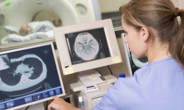
High-field and Hybrid
European Hospital met up with Professor Harald H. Quick, PhD, who was appointed Director of the Erwin L. Hahn Institute (ELH) for Magnetic Resonance (MR) Imaging this February.

Being in charge of highfield and hybrid magnetic resonance (MR) imaging, Quick shared his views on the status of and the imminent research into MRI systems operating at 7.0 Tesla (7T) as well as the combined positron emission tomography (PET)/MR hybrid imaging technology.
7 Tesla MRI is a platform for clinically-oriented research, but not yet a medical product
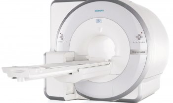
MR imaging systems at 7T magnetic field strength or above are by now an established platform for clinically-oriented research. According to Quick, there are currently about 50 such systems installed worldwide. They were built to enable highfield MR imaging of the entire human body. 7T MRI technology is “the carrot stick”, explains Quick, “which can be used to obtain more details than at 1.5T or 3.0T due to its inherent high signal-to-noise-ratio”. However, Quick reckons that “7T is still about two or three years away from becoming a medical product, as issues such as safety and standardisation of the technology have not yet been sufficiently addressed”.
The inherent advange of this technology lies in its excellent soft-tissue contrast, and high spatial resolution which permits to better determine “the anatomical structure down to the finest details“ , summarizes Quick, who also points out that 7T “enables to further characterize the structure of lesions in multiple sclerosis rather than just counting them. Ultimately, Quick hopes that, for example, depicting tumour vascularisation in a superior way will empower clinicians to pick up structural changes at a much earlier point in time and therefore raise the likelihood of improving patient outcome.
Quick points out that beyond displaying fine structures, depicting tissue and organ function plays an increasing role in highfield MRI. Due to the increased sensitivity of highfield MRI the blood-oxygenation-level-dependent (BOLD) effect detectable during a functional MRI, “7T allows to register increased oxygen consumption in the brain, which means that we can indirectly watch the brain think”. But for this to happen, just using strong magnetic fields is not enough. So called radiofrequency (RF) coils, or antennas, are imperative to excite and readout the MR signal from the particular body area to be investigated. As RF coils for the head are already commercially available, the clinical research into the use of 7T technology focuses mainly on imaging the brain. However, imaging the rest of the body is currently a piecemeal attempt with different institutes concentrating on designing RF coils for specific body parts, such as heart, spine or extremeties. Institutes do this in collaboration with MR system producers and clinicians.
ELH in Essen, Quick reveals, “has been devoted to a holistic approach, aiming at depicting each part of the body with specifically designed RF coils: Our group is internationally positioned to develop high-frequency RF transmitter and receiver coils plus multi-channel systems”. In the field of 7T highfied MRI Quick’s team’s research focuses on:
• imaging the heart, prostate and mammography
• designing RF coils and RF technology for whole-body imaging
• ensuring the safety of those RF coils
• adapting sequences for clinical highfield MR imaging.
PET/MR is closer to the clinical side than MRI at 7 Tesla
Having worked closely with Siemens Healthcare on the introduction and advancement of PET/MR hybrid imaging during his tenure as professor at the Institute of Medical Physics at the Friedrich-Alexander-University in Erlangen-Nuremberg, Quick brings to his new job a wealth of experience in the PET/MR realm along with excellent contacts to the industrial partners as well as the PET/MR imaging community.
Status of PET/MR
Quick estimates that globally there are currently about 50 installed PET/MR systems. Unlike 7T highfield MRI, PET/MR systems are already labeled as medical products and posses more extensive clinical experience. However, reimbursement and refinancing are still unresolved, as is their use case, which still needs to be defined.
Will PET/MR cannibalise PET/CT?
Quick does not believe that PET/MR will cannibalise or even replace PET/computed tomography (CT): “Both modalities will coexist. Factors such as availability and costs will play a major role, especially with PET/MR being time and cost intensive”. It will require a careful assessment of both modalities’ strengths and weaknesses to determine their best use cases. Current research suggests that PET/MR is particularly promising as a diagnostic imaging modality in whole-body oncology (for depicting soft tissue tumours), pediatric oncology (due to the reduced amount of ionizing radiation when compared to PET/CT) and neurology (combining excellent soft tissue contrast with high sensitivity).
Future areas of PET/MR research at ELH
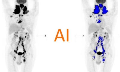
Quick is passionate about fostering interdisciplinary PET/MR research, joining the efforts of clinicians, manufacturers, physicists and other scientists. Asked about his immediate research priorities, Quick comes up with a long laundry lists of projects. He is particularly keen to investigate the potential for reducing radiotracer due to the high sensitivity of the PET detectors in the context of PET/MR, explore ”motion correction technologies to correct for breathing and cardiac motion in view of the relatively long PET data acquisition times”, work on attenuation correction (AC) and to develop MR sequences, which will help to provide bone information for MR-based AC. Furthermore, Quick wants to optimise the hybrid imaging workflow in such a way that it “maximizes diagnostic information while minimizing acquisition time”.
It is certainly an ambitious list, but Quick is optimistic: “There is a lot of research waiting for us , but the team is enthusiastic and with 7T highfield MR and hybrid PET/MR we have two of the latest and greatest tools for MR imaging research available here in Essen”.
Profile:
Professor Dr Harald H. Quick studied biomedical and physical engineering at the University of Applied Sciences, Aachen, Germany. Following his first professional appointment as Research Associate at the MRI Centre of the University Hospital Zurich, Switzerland, in 1999-2000 he spent a research year at Johns Hopkins University in Baltimore/USA. Back in Germany, he joined the University Hospital Essen, where he founded MR-Innovation GmbH. From 2006 to 2009 he was Managing Physicist at Erwin L. Hahn Institute for MRI. In 2009 he was appointed Professor for MRI at Friedrich Alexander University
06.03.2014