ShearWave Elastography enters new applications and medical fields
In 2005, when they founded the French ultrasound company SuperSonic Imagine, Jacques Souquet PhD and Claude Cohen-Bacrie had to convince radiologists that they not only offered a new product but also a completely new ultrasound technology that could measure entirely new parameters.
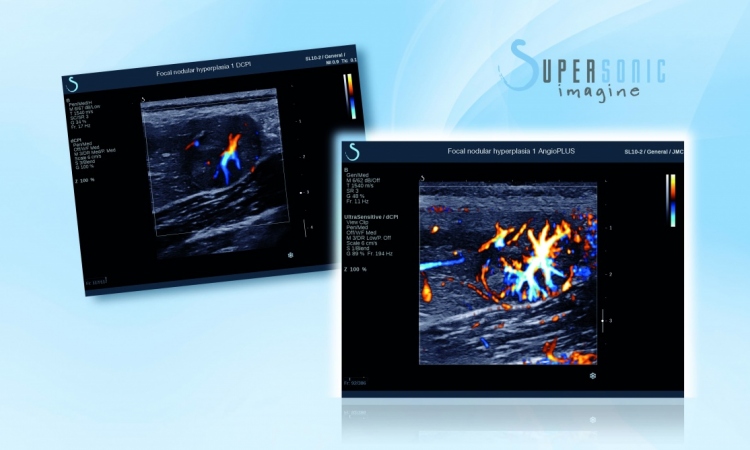
This was ShearWave elastography, which measures tissue stiffness in kilopascals, an additional tool to complement gray scale for the characterisation of tissue. They decided it would initially provide the most benefits in breast cancer assessment. Within six years this has been well proven and ShearWave elastography has spread to other clinical usage. In an interview with European Hospital during RSNA 2010, the firm’s COO and CSO Claude Cohen-Bacrie outlined what ShearWave Elastography already provides as well as future expectation.
‘The initial idea of positioning ShearWave elastography in breast diagnosis has been to improve the accuracy of BIRADS classifications. We were interested in knowing if adding features such as the shape and stiffness of the shear wave image of the lesion, on top of the greyscale information would improve diagnostic accuracy. After a two-year multicentre clinical trial, involving 1,800 patients, we know it is the case: We showed that, due to ShearWave elastography, a relevant number of BIRADS 4a cases could be downgraded to BIRADS 3, and vice versa. The impact on therapy management is obvious as this reduces false positives and false negatives. Based on these finding, we later launched a linear volumetric transducer that enables the user to assess lesions in 3-D, with targeted applications such as therapy planning or monitoring. Measuring the stiffness of a lesion can indeed potentially provide better assessment of tumour size or be helpful information about the success of a type of therapy.
‘The next challenge we face is breast cancer screening. As it is often difficult to image dense breasts with mammography, we have seen that ultrasound can image them very well and without any risk from radiation exposure. Studies have already shown that ultrasound can find more cancers compared with mammography alone but, the false-positive rate remains an issue. We demonstrated that ShearWave elastography is resolving that high false positive rate, allowing ultrasound to become economically viable for screening.’
‘This development in the breast imaging sector perfectly reflects our overall strategy of answering the questions: How does the technology we develop apply to the organs we want to image and, how does this technology can become a clinical innovation?
‘Another good example is the characterisation of thyroid nodules. We are seeing more and more imaging of the thyroid because nodules are a frequent problem with people over fifty. Almost half of this population has thyroid nodules. Today, the selection and classification with common ultrasound is not as standardised as in mammography. In addition, now-a-days the specificity of Fine Needle Aspiration (FNA) is low. So, looking at our experiences in breast ultrasound, we are convinced that ShearWave elastography can improve the pre-selection and classification of thyroid nodules resulting in better FNA outcomes.
‘Likewise, in liver assessment, where we offer a tool for imaging the stiffness of the liver, this could potentially help in selecting the right candidates for liver biopsies. Furthermore, in liver therapy monitoring ShearWave elastography has an important role to play to evaluate the reaction of the liver to the therapy. For example, ShearWave elastography can locate exactly where the Radio Frequency Ablation was induced as the stiffness of tissue increases with this treatment.
‘Finally, a question we are currently working on is: Could ShearWave elastography improve the diagnosis of prostate cancer? Today, the Gold Standard is to launch a certain number of ultrasound-guided core biopsies, unfortunately this technique incurs a significant number of false-negative results. This technique could be aided by using ShearWave elastography to measure the stiffness in the prostate and used to help guide the biopsy in order to achieve better samples.
‘So, step by step, and through our expanding experience, ShearWave elastography is entering new applications and fields such as screening, diagnosis and therapy monitoring of several organs.’
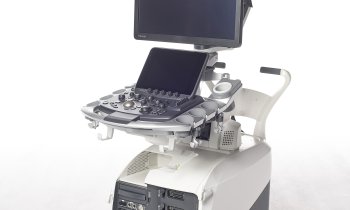
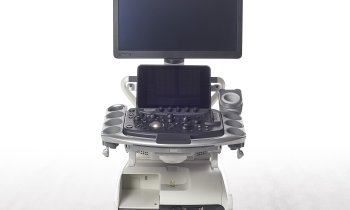
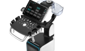
Supersonic Imagine is in Booth 4, Extension Expo A
ECR 2011 symposium
4 March. 12 -13.30. Room G/H
Exploring breakthroughs against cancer and fibrosis
Claude Cohen Bacrie, moderator and speaker on the Presentation of Clinical Benefits of ShearWave elastography Worldwide Breast Trial: Model and Final Results.
Other speakers: Dr Schäfer (TBC) on Validation of ShearWave Elastography Worldwide Breast Trial Model with European Cases; Prof. Correas on Preliminary experience with ShearWave elastography in the management of prostate cancer, and Dr Ferraioli on ShearWave elastography for the evaluation of liver fibrosis.
02.03.2011
- breast cancer (544)
- elastography (89)
- imaging (1488)
- medical technology (1447)
- prostate cancer (195)
- tissue (193)
- ultrasound (721)